Digital Dental X-Ray
Latest in X-Ray technology, highest performance and precision combined with lowest radiation dosage
Digital Dental X-Ray
Latest in X-Ray technology, highest performance and precision combined with lowest radiation dosage
12 TYPES OF IMAGING
Taken with strictest possible safety measures that protect whole body, reproductive organs, and thyroid gland.
PROFESSIONAL STAFF
X-rays are taken, edited, and archived by our professional team of dedicated radiologic technicans.
LATEST TECHNOLOGY
The latest digital dental X-ray technology we are using emits upwards of 90% less radiation than older X-ray machines.
X-RAY IMAGES
All X-rays are immediately archived in electronic format and they are available for transfer on CD or via email.
Polyclinic Šlaj-Anić provides services of the most modern digital dental X-ray in Croatia. Our Planmeca ProMax 3D Mid system belongs to the latest generation of dental diagnostic technology marked by Scandinavian manufacturing quality, high precision and low radiation dosage.
ProMax 3D Mid system produces digital X-ray diagnostic images of highest quality and precision. Diagnostic palette covers no less than 12 different types of imaging. All images are taken while following strict safety rules and procedures, including protection of patient’s whole body, genitals, and thyroid gland. Imaging and editing is done solely by professional radiologic technicians.
X-rays of teeth and jaws are diagnostic tools for analyzing anatomical characteristics of teeth, condition of bone in periodontitis, diagnosing pathological processes (tooth decay, granulomas) and determining thickness of the bone for implant planning.
We conduct X-ray cephalometric analysis as the essential diagnostics for all our orthodontic patients. We also offer the service of cephalometric X-ray analysis to our out-colleagues, specialists in orthodontics.
Digital images are faithfully displayed immediately on the screen and are then processed by the specialised software for a more precise analysis: enhancement of certain details, adding contrast, and changing focused sections. We are immediately able to directly store digital data and we can also send images electronically to third parties. The most important advantage of our digital dental X-ray imaging technology is the health benefit. Our modern digital dental X-ray device reduces the radiation dose for the patient up to 90% (proven and tested average is 77%).
As mentioned before, digital dental X-ray used in Polyclinic Šlaj-Anić belongs to the latest generation of digital X-ray imaging devices. It is far superior to older technology and provides higher quality and precision in diagnostics while decreasing radiation dosage.
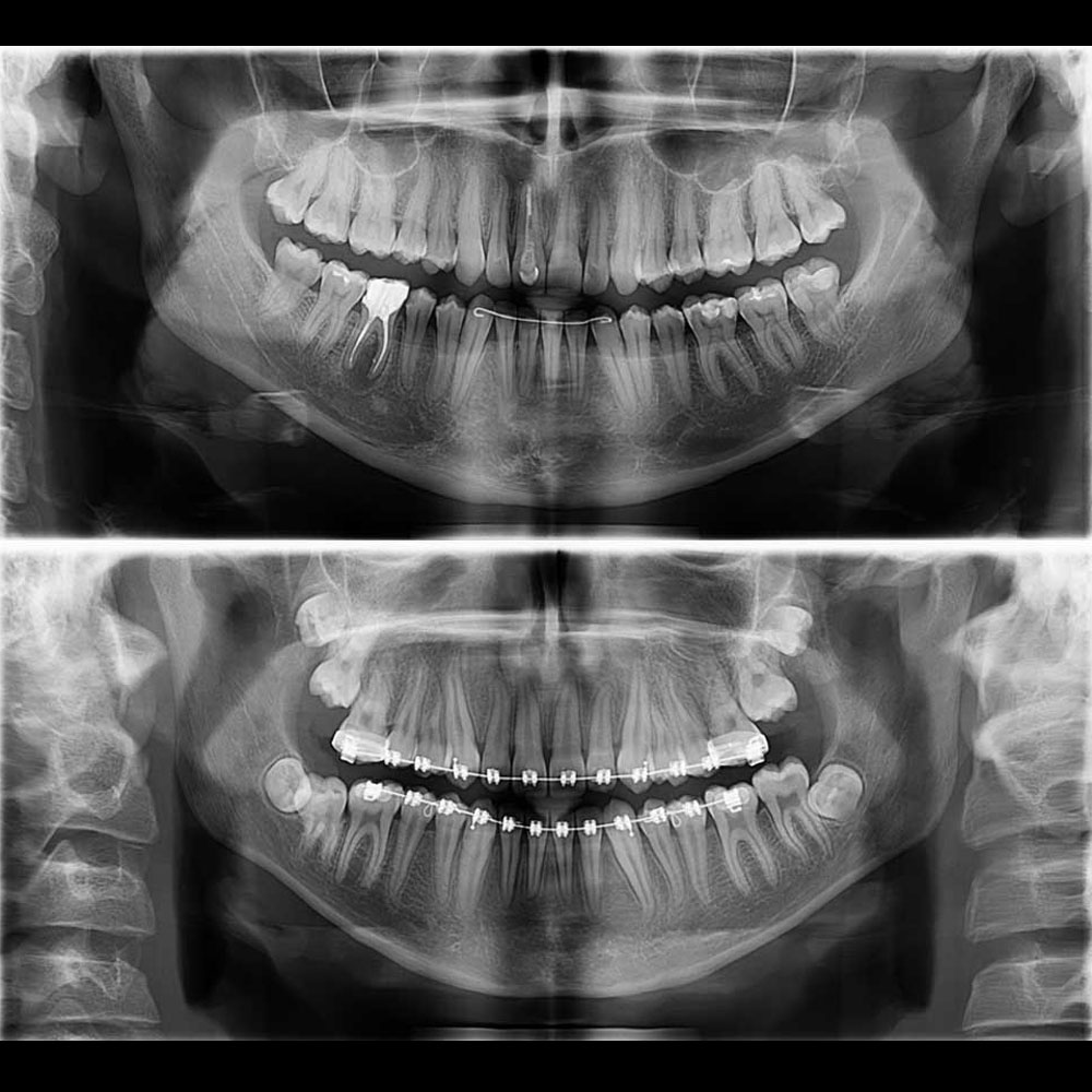
PANTOMOGRAM
Dental panoramic radiograph covers upper and lower jaw, teeth, temporomandibular joints and associated structures.
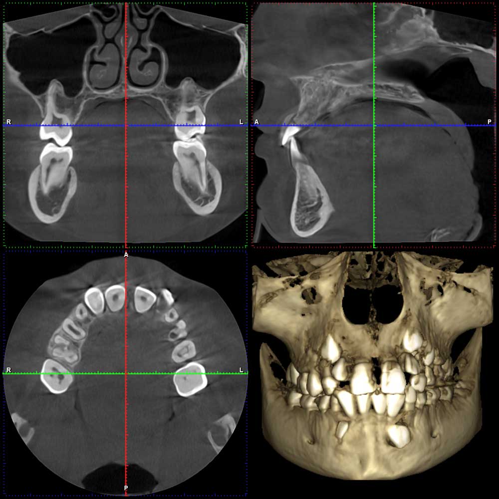
CBCT
Cone-Beam Computer Tomography is 3D image of both jaws, indivudual teeth, joints and maxillary sinuses .
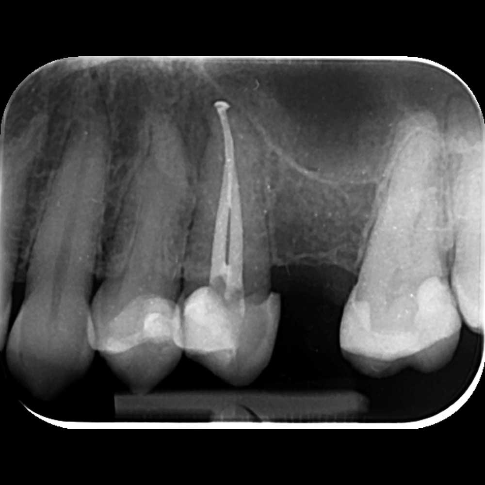
INTRAORAL IMAGING
Intraoral periapical radiograph focuses on detailed imaging of individual tooth and its surrounding structures.
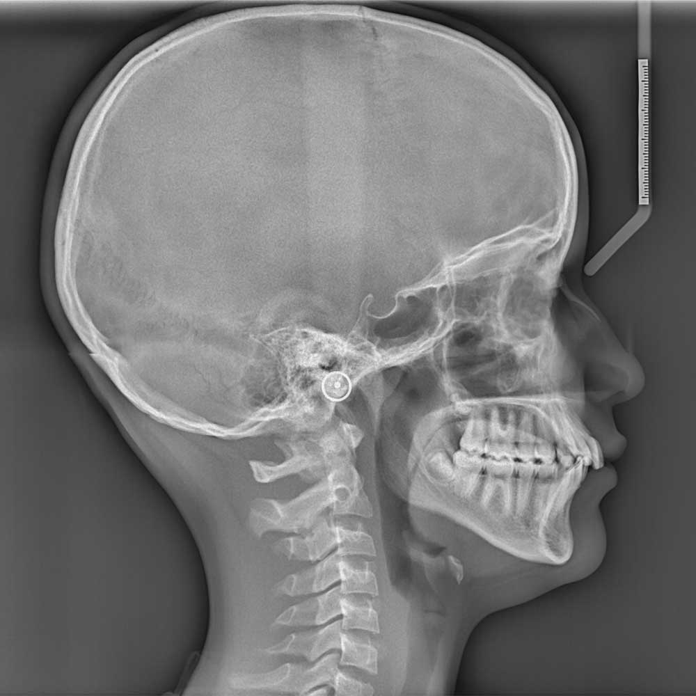
CEPHALOGRAM
Lateral craniogram (LL) is a side image of the head, all of the head and neck bones are captured in profile.
Planmeca ProMax 3D Mid
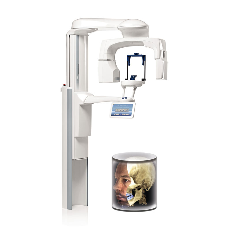
Since its founding Polyclinic Šlaj-Anić was equipped with high quality dental X-ray system. Our digital dental X-ray has been modernised and upgraded in april of 2018. with the arrival of Planmeca ProMax 3D Mid system. In addition to panoramic, cephalometric and 3D CBCT imaging this system has the ability to digitally integrate all of them together.
This is the latest in medical technology of digital imaging and created specifically for the use by doctros of dental medicine and oral and maxillofacial surgeons. High-quality diagnostic data is obtained with minimal radiation dosage.
3D imaging is necessary in diagnostics of various ailments affecting tooth, jaws, and surrounding structures. It is also crucial for planning of complex medical procedures like dental implant placements, wisdom tooth extraction, dental surgery, extraction of impacted teeth etc.
CBCT technology enables 3D imaging of whole jaw, or we can limit the imaging field to just a few teeth. We can also take highly precise CBCT images of temporomandibular joints and maxillary sinuses. 3D CBCT tooth imaging on Planmeca ProMax 3D Mid system enables limitless number of possible diagnostic combinations.
Soredex-Minray
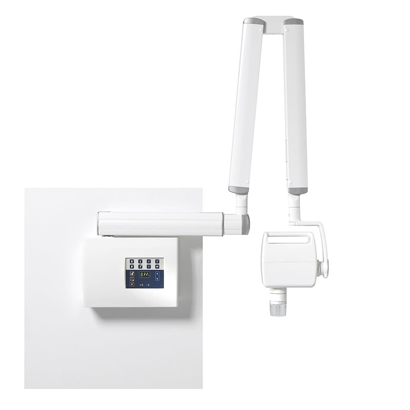
For intraoral imaging we use Soredex-Minray digital X-ray and intraoral RVG sensors designed to operate in pair with scanner Digora Optima New. These sensors give us a better and sharper focus. Eeach image can be processed so that every tooth root is clearly visible.
Depending on the medical need we use the following types of X-ray imaging:
- intraoral imaging of single tooth (small images of individual teeth)
- pantomogram (panoramic X-ray image)
- segmental pantomogram (left or right quadrant, frontal)
- pantomogram of children (specially reduced radiation dose)
- pantomogram 1: 1
- lateral craniogram different projections: LL, PA, AP
- bite wing imaging
- occlusal imaging
- CBCT of desired areas: maxillary sinuses, temporomandibular joints, individual teeth, upper and lower jaw, nerve canals etc.
- imaging of temporomandibular joints
- imaging of maxillary sinuses

